The anatomy of the human body is intricate, and your eye is no different. There are a number of parts in your eye that are essential to how they function and allow you to see the world around you. So, how do they work?
Let’s discuss how your eyes function, the parts of your eye and what they do, and common conditions that can affect your eye’s ability to work.
Table of Contents
How Do Your Eyes Work?
Your eye structure lets light enter and pass through a series of clear components and sections. These structures bend and focus light, adjusting how far the light beams travel before they come into focus. Because the focus needs to be so precise, your eye muscles make subtle changes to the shape of your eye, moving the focus point so it lands correctly on the retina.
When light lands on the cells of your retinas, those cells send signals to your brain. The signals describe everything about the light, including the color, how intense it is, and other details. Your brain then processes these signals and uses them to create the image you see.
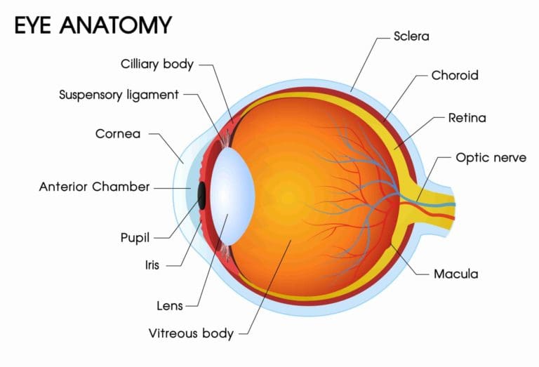
Parts of the Eye and Their Function
Cornea
The cornea is the transparent circular part in the front of the eyeball. It refracts light that enters the eye onto the lens, which then focuses it onto the retina. The cornea doesn’t contain any blood vessels and is very susceptible to pain.
Sclera
The sclera is the white part of the eye, which acts as a tough cover surrounding the iris.
Conjunctiva
The conjunctiva is a thin, clear membrane that protects your eye. It covers the inside of your eyelid and the white of your eye and works with your glands to create tears and protect the white part of your eye from damage.
Aqueous Humor
The aqueous humor is the clear liquid inside the front part of your eye. It nourishes your eye and keeps it inflated. Your eye constantly produces a small amount of aqueous humor while an equal amount flows through the trabecular meshwork at the drainage angle.
Iris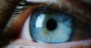
The iris is the colored part of your eye that helps regulate the amount of light that enters your eye. When there’s a bright light, the iris closes the pupil to let in less light. When the light is low, the iris does the opposite and opens up the pupil to let in more light.
Pupil
The pupil is the dark center opening in the middle of the iris. The pupil changes size to adjust for the amount of light available. They become smaller in bright light to let less light in and open more in low light to allow more light inside.
Lens
The lens focuses light rays onto your retina. The lens is transparent and, if necessary, can be replaced with intraocular lenses. Your lens deteriorates as you age, which can result in the need for reading glasses.
Vitreous Humor
The vitreous humor is the clear, gelatinous substance that fills the central cavity of the eye.
Retina
The retina is a light-sensitive layer that lines the interior of your eye. It’s composed of about 125 million rods that help you see in dim light and between 6-7 million cones that help your eyes function in bright light.
Macula
The macula is a yellow spot on the retina at the back of your eye. It surrounds the fovea.
Optic Nerve
The optic nerve is a bundle of more than a million nerve fibers that carry visual messages from the retina to the brain. This is necessary because your brain controls what you see by combining images. The retina sees images upside down and the brain turns images right side up.
External Muscles
External eye muscles are responsible for moving the eye along three axes. They move horizontally, which is toward or away from the nose. They also move vertically, which is either elevation or depression, and torsional, which brings the top of the eye toward or away from the nose.
There are seven extraocular muscles:
- Levator palpebrae
- Superioris
- Superior rectus
- Inferior rectus
- Medial rectus
- Lateral rectus
- Inferior oblique
- Superior oblique
Common Eye Conditions and Disorders
Refractive Errors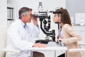
A refractive error is when the natural shape of your eyes makes your vision blurry. This could be caused by the shape of your eyeball, cornea, or lens making it difficult for your eye to focus correctly on objects that you’re looking at.
There are three main refractive errors:
- Nearsightedness: you have difficulty seeing distant objects, but can see objects that are close to you clearly.
- Farsightedness: you have difficulty seeing objects that are close to you clearly. This is the opposite of nearsightedness.
- Astigmatism: you have difficulty seeing any objects at any distance without them being blurry. This generally occurs when your eye is shaped like a football, making light that enters your eyes bend and distort more than it should.
Corneal Disorders
Corneal disorders refer to a number of conditions that affect your cornea.
There are the three most common:
- Keratitis: this occurs when there’s inflammation in your cornea that can either be infectious or noninfectious. Bacteria are the most common cause of infectious keratitis, while noninfectious keratitis can be caused by eye injuries or other conditions that dry out your eye.
- Corneal Ectasia: this is a group of conditions that change the shape of your cornea, causing it to thin and bulge outward.
- Corneal Dystrophy: this is a group of genetic disorders that involve abnormal deposits of proteins, fluid, and other materials in one or more layers of your cornea.
Retinal Disorders
Retinal diseases affect your retina, the back layer of your eye, and often cause symptoms that affect your vision. If they’re not treated quickly, they may eventually cause blindness or low vision.
Some examples of retinal disorders include:
- Retinal Detachment: this occurs when the retina pulls away from the tissues supporting it, causing flashes and darkening side vision.
- Diabetes-Related Retinopathy: this is a condition that weakens the blood vessels in the retina. Anyone with diabetes is vulnerable to this disorder and it can lead to vision loss or blindness.
- Age-Related Macular Degeneration: this is an age-related condition that affects your macula and causes you to lose your central vision.
Optic Nerve-Related Conditions
Damage to your optic nerve can cause vision loss that affects one or both of your eyes.
Some nerve-related conditions include:
- Glaucoma: this is a group of diseases that occurs when the fluid pressure inside your eye slowly rises and damages the optic nerve.
- Optic Neuritis: this occurs when the optic nerve is inflamed. It can trigger infections and immune-related illnesses.
- Optic Nerve Atrophy: this is a condition in which there is damage to the optic nerve, which can include poor blood flow, disease, trauma, or exposure to toxic substances.
Age-Related Eye Disorders
Age-related eye disorders and conditions are one of the primary causes of blindness and low vision in the United States.
These conditions include:
- Cataracts: this is the clouding of your eye lens that is the leading cause of vision loss in the United States.
- Dry Eyes: this is a condition that happens when your tear glands can’t make enough tears or produce poor-quality tears, drying out your eye.
- Floaters: these are tiny spots or specks that float across your field of vision.
Common Signs of Eye Conditions
If you notice any of these symptoms, they can be a sign that you have an eye condition:
- Irritation
- Red eyes
- Watery eyes
- Discharge coming from your eyes
- Trouble seeing clearly
- Decrease or loss of vision
- Disrupted vision
- Light sensitivity
Eye Treatment With Cleveland Eye Clinic
If you notice any of the signs of an eye disorder, schedule an appointment for a comprehensive eye exam with the knowledgeable professionals at Cleveland Eye Clinic. Our reliable team is committed to ensuring that you get the very best treatment and results.
We also offer premier treatments and procedures, like LASIK, to give you the vision that you’re happy with.
Your eye health is essential to your overall health. Reach out to us today.